How to find the nub on your scan (and read it)
The nub — the genital tubercle — sits at the very base of the spine, between the legs, on a true side-profile scan around 12 weeks. Trace the spine down to the bottom and look for a small bright bump at the end. Its angle to the spine is the whole game: over 30° leans boy, roughly parallel leans girl.
Quick answer
- Where: the tail-end of the spine, between the legs, as a small raised point.
- What: the genital tubercle — the bud that becomes penis or clitoris.
- When it's visible: a clean mid-sagittal (side) profile at 12–14 weeks.
- How to read it: angle to the spine — over 30° boy, under 30° girl.
- If you can't find it: the image probably isn't a true side profile.
What the nub actually is
Every fetus starts with the same structure — the genital tubercle, a small bud between the legs. In boys it lifts and elongates into the penis; in girls it stays lower and becomes the clitoris. In the first trimester, before that difference is obvious, the tubercle is simply "the nub." Nub theory reads the angle it makes with the spine, because the male and female versions rotate along different paths as they develop. For the full method and its accuracy, see nub theory explained.
Step by step: where to look
- 1. Confirm it's a side profile. You need a mid-sagittal view — the spine should run as a clean, continuous line along one edge of the image. No spine line, no valid read.
- 2. Follow the spine to the base. Run your eye from the head down the spine to the tailbone end. The nub is right there, at the bottom, between the legs.
- 3. Find the small bright bump. The genital tubercle shows up as a short, raised point of tissue — brighter than the surrounding fluid.
- 4. Separate it from the legs and cord. The thighs and the umbilical cord sit nearby and love to confuse the picture. Make sure the bump you're looking at is the tubercle, not a cord loop.
- 5. Check the week. The nub is only readable from about 12 weeks. Earlier, boys and girls look nearly identical.
How to measure the angle
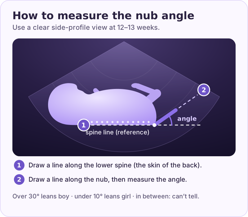
Want to try it on your own scan? Our free nub angle calculator lets you place the two lines on your photo and measures the angle. The photo never leaves your device.
Draw one line through the lower spine and a second line along the nub. The angle between them is the genital tubercle angle:
- Boy: the nub tilts up and away — 30° or more.
- Girl: the nub lies flat and parallel — under 30°.
- Borderline (roughly 20–29°): genuinely inconclusive; don't force it.
The 30° threshold comes from the original nub study (Efrat 1999). For the deep dive on the measurement itself, see the angle of the dangle.
Found your nub? Upload the scan to the nub guesser and let Peek take a look. The full angle read happens in the app.
Open the nub guesser →Why people can't find the nub
- Wrong plane. A front-on or tilted image doesn't show the tubercle's angle — often it hides the nub entirely.
- Legs in the way. Crossed or tucked legs cover the exact spot you need to see.
- Too early. Before 12 weeks the tubercle hasn't developed a readable angle.
- Cord confusion. A loop of umbilical cord between the legs is the classic false "nub."
- Low resolution. A compressed phone photo of a scan can blur the tubercle into the background.
If none of your images are a clean side profile, the most honest conclusion is that this scan can't be read — not that it's a girl because the nub "looks flat."
FAQ
Sources
- Efrat Z, Akinfenwa OO, Nicolaides KH. First-trimester determination of fetal gender by ultrasound. Ultrasound Obstet Gynecol. 1999;13(5):305–7. PubMed
- Efrat Z, Perri T, Ramati E, Tugendreich D, Meizner I. Fetal gender assignment by first-trimester ultrasound. Ultrasound Obstet Gynecol. 2006;27(6):619–21. PubMed
- Alfuraih AM et al. The predictive accuracy of anogenital distance and genital tubercle angle for first-trimester fetal sex determination. Diagnostics (Basel). 2024. PubMed
Written by the BabyPeek editorial team from the studies above. How we research and write. For entertainment, not medical advice.
Let Peek measure it for you
Not sure you've spotted the right bump? BabyPeek lets you mark the nub and spine on your uploaded 12-week scan and measures the angle on YOUR image — then combines it with four other methods for a majority verdict and a shareable reveal.
For entertainment only. This is not medical advice — confirm your baby's sex with your doctor's anatomy scan.